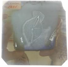
And I would also add, everyone has beauty, but not everyone can see it (“…mirror mirror on the wall who is the fairest of them all?” - See the picture below of our Volunteer Co-ordinator, Ali Thomas, trying to take some pictures of a lantern slide).
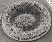
The diatoms are indeed unique organisms that come on an array of amazingly beautiful shapes, forms, structures and/or with extraordinary ornamentations on their walls, sometimes quite complex. To be able to see this and admire them, it is needed (unfortunately) at least some magnification; perhaps this is their price, for being such striking organisms. The more I look at them, and learn about them, the more I discover their beauty.
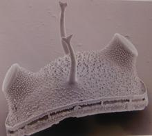
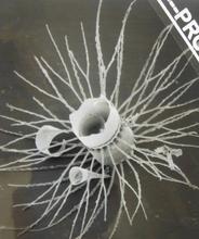
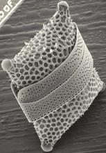
As Goethe once stated, “Beauty is a manifestation of secret natural laws, which otherwise would have been hidden from us forever” Thanks to the microscopes, or nowadays, to the SEM (Scanning Electron Microscopy), the beauty of diatoms and all their details are exposed. The SEM (which started to become available from 1960's, more precisely, 1965) has revolutionised science, and it has been also very useful for Diatoms, because it has helped to clarify so many questions that the microscopes were not able to answer, in terms of morphological distinctiveness on those times.
Much before SEM, there were microscopes and lantern slides, or glass plates (as the example pictured above). The lantern slides arrived 10 years after the invention of photographs; the lantern slides could be projected so many could see at the same time. They were common until 1950's then their use (and production) declined, and after that, came the 35mm microscope slides....so integral to this current V Factor collaboration.
The current project, focusing on T. Comber diatom slides (under the V Factor initiative) will try to expose the beauty of numerous diatoms that have been identified and listed by T. Comber from a series of ca. 3000 slides sampled worldwide. Most of the images will be those that have been taken (by T. Comber) under usual microscopes available on those times (early 1900's)- see image below. However, they don’t diminish their beauty or even their importance. Although one could, if time and resources are on hand, try to re-photograph them, under a much better microscope or in some cases, as we have some of the samples (the bottle collection) from which the slides were made, prepare the material for SEM.

If you would like to see more of the exquisite and beauties from the microscopic world of the Diatom , visit us on Thursdays, in the Specimen Preparation Area (SPA) of the Darwin Centre at the Natural History Museum.
Comments
Making the invisible visible; Thomas Comber diatom sample study
Hello Dr. Jovita,
I am interested in the continued data provided from the study of the millions of Diatom species during Dr. Comber's original sample study in the 19th Century. So fascinating these phytoplankton. I wonder if you might answer a question for me or refer me to deeper research or studies on line. Has there been any data that would relate most Diatoms from the earth to the bacteria commonly known as blue-green-Algae or lab name of Cyanobacteria? It would be a great service to many who have questioned. Your work (Dr Comber's) work would provide more scientific detail since you have so closely studied most species.
Add new comment